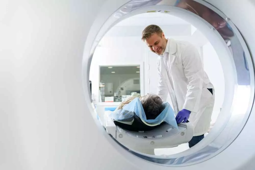
Modern imaging techniques in neurological diagnosis and psychiatry
Neurological diagnosis and psychiatry are medical fields that have undergone tremendous advances over the past few decades. Modern imaging techniques allow a more precise assessment of brain structures and the study of various processes in the body, which has improved the diagnosis and treatment of many neurological and psychiatric diseases.
Magnetic resonance imaging (MRI)
Magnetic resonance imaging (MRI) is one of the most important imaging techniques used in neurological and psychiatric diagnosis. MRI produces detailed images of brain structures using a strong magnetic field and radio waves. This non-invasive method allows assessment of both anatomical structures and brain function.
In neurological diagnostics, MRI is used to detect and monitor brain changes associated with diseases such as stroke, cancer, multiple sclerosis and Alzheimer's disease. In psychiatry, MRI allows assessment of structural and functional brain changes in patients with psychiatric disorders such as depression, schizophrenia or bipolar affective disorder.
Computed tomography (CT)
Computedtomography (CT) is another popular imaging modality used in neurological diagnosis. CT produces fast and precise 3D images of the brain using X-rays. This technique is particularly useful in detecting structural changes, such as tumors, hematomas or brain injuries.
In psychiatry, CT is used less frequently than MRI, but can still provide valuable information about possible brain lesions in patients with psychiatric disorders.
Electroencephalography (EEG)
Electroencephalography (EEG) is a technique for recording the electrical activity of the brain. It is a non-invasive procedure in which electrodes are placed on the patient's scalp. Brain waves invisible to the eye are recorded, which can provide information about brain activity and possible abnormalities.
EEG is commonly used in the diagnosis of epilepsy and other seizure disorders. It can also be used to diagnose certain mental illnesses, such as schizophrenia and mood disorders.
Positron emission tomography (PET) imaging
Positron emission tomography (PET) imaging is a technique that assesses brain function by imaging the distribution of radiation emitted by radioactive substances (labeled tracers) introduced into the patient's body. PET provides information on blood flow, metabolism and receptor density in different areas of the brain.
In neurological diagnostics, PET is particularly useful in evaluating healthy aging brains and in detecting various neurodegenerative diseases such as Parkinson's disease, amyotrophic lateral sclerosis and Alzheimer's disease. In psychiatry, PET can provide information on functional brain activity in patients with mental disorders such as depression, schizophrenia or addiction.
Summary
Modern imaging techniques such as MRI, CT, EEG and PET are revolutionizing neurological diagnosis and psychiatry. They allow accurate assessment of brain structures and function, which contributes to rapid and accurate diagnosis of many neurological and psychiatric diseases. This also makes treatment more effective and targeted. It is worth paying attention to these advanced imaging methods and using them to provide patients with the highest possible level of medical care.

Add comment